")
Q&A
StemJournal posed some questions to Martin and below you can read his answers:
Q: What are the key takeaways from this review?
A: The main take home point is that there are many questions on how stem cells work and that is why it is critical that we track stem cells after delivery. By combining tracking of stem cells in vivo with functional assessment of the function of the organ under study, we will gain a better sense of the efficacy of these therapies. Also, that there is no “one size fits all” when it comes to imaging, all depends on the research question posed.
Q: In general, how do you feel the molecular imaging techniques in the field have changed in the last 10 years since the original chapter review was published in StemBook?
A: Since the first publication of this review, several advances have occurred. New modalities have appeared, for example, magnetic resonance reporter genes and optoacoustics, and there have been improvements in modalities that already existed (for example, fluorescence tomographic imaging).
Q: What was your approach to writing the review for StemJournal? How did you decide which sections to focus on for the update?
A: As a clinician – I am a cardiologist – I always have the clinical application in mind. Thus, we focused on modalities that have a potential for clinical application, such as magnetic resonance (MI), positron emission tomography (PET), or single photon emission computed tomography (SPECT), or can be adapted to clinically applicable modalities, for instance bioluminescence.
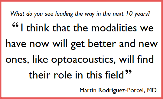
Q: What have the significant advances been, and what do you see leading the way in the next 10 years?
A: I think that the modalities we have now will get better and new ones, like optoacoustics, will find their role in this field. In the next few years, we will be learning more and more about how cells work and what we can do to make them more efficacious as a therapeutic strategy.
Q: How does the content of the paper(s) relate to your research, and what is your current/future focus in this field?
A: This field is the main area of interest in my laboratory. My focus is to try to understand what happens with stem cells after they are delivered, especially for cardiovascular applications. I expect to continue to work in this field in the next few years, as I am convinced that we need to work hard to advance these therapies. I have been fortunate to work with leaders in the field (Dr. Wu, Dr. Gambhir) and I want to put all the effort I can to advance knowledge in this field.
Q: Anything else you’d like to share?
A: I am convinced that regenerative therapies have a role in health care and is up to us to find what it is. There are many questions remaining on the stem cell field, questions in which in vivo imaging will play a significant role in the future. We should not be discouraged by some of the results of stem cell studies. In fact, it further highlights the important role that imaging will play on this field as we go forward.
--------
Thank you to Martin for answering our questions! The StemJournal article can be viewed here, and it will be co-published on StemBook as the revised chapter soon (the original chapter on StemBook can be viewed here.).

Calling all authors who have previously published chapters on stembook.org. If you feel a revision of your previous article is due, please submit your updated manuscript to StemJournal. See here for full guidelines, or contact us if you have queries via: stemjournal@iospress.com

