Loading...

HSCI Retreat 2020 Abstract 5

Organoid Maturation by Circadian Entrainment

Juan R. Alvarez-Dominguez,*1 Julie Donaghey,1 Niloofar Rasouli,1 Jennifer H. R. Kenty,1 Aharon Helman,1 Jocelyn Charlton,1,2 Juerg R. Straubhaar,1 Alexander Meissner,1,2 and Douglas A. Melton1 
1 Department of Stem Cell and Regenerative Biology, Harvard Stem Cell Institute, Harvard University, Cambridge, MA, USA
2 Department of Genome Regulation, Max Planck Institute for Molecular Genetics, Berlin, Germany

* Presenting and corresponding author: juanralvarez@fas.harvard.edu

Submitted: Jun 11, 2020; Published online: Jul 27, 2020


ABSTRACT

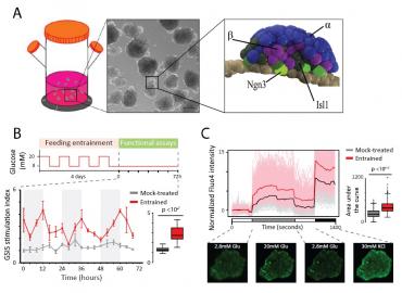
Stem cell-derived organoids that recapitulate endogenous physiology could transform disease research and therapy, yet most methods yield products that function like fetal, not adult tissues. Organoids are typically grown in constant environments, whereas our tissues turn functionally mature in the presence of daily physiological rhythms. To better understand maturation-driving mechanisms, we studied epigenome dynamics during the stepwise formation of pancreatic islet organoids from human pluripotent stem cells (Fig.1A) (Alvarez-Dominguez et al., 2019, Cell Stem Cell, 26, 108–122 e110). We devised tools to purify intermediates across islet lineage progression, and profiled their DNA methylation, accessibility, and histone modification changes. Unexpectedly, we found BMAL1/CLOCK footprints enriched at genomic sites activated as organoids gain glucose-responsive insulin secretion. Moreover, we uncovered transcriptional loops between circadian effectors DEC1/2 and factors linked to mature insulin responses. This suggested circadian clocks may foster organoid maturation. Indeed, entrainment to 12h feeding-fasting cycles (Fig.1B) induces organoid clocks, eliciting pulsatile synthesis of energy metabolism and insulin secretion effectors, including antiphasic insulin-glucagon pulses. This triggers metabolic rhythms and cyclic insulin responses with higher glucose threshold and single-cell secretion (Fig.1B,C), a hallmark of islet maturity. Clock-entrained organoids gain stable epigenetic changes at genes enabling mature insulin responses, and function within days of transplant. Clock control of islet maturity effectors depends on Dec1 binding their promoters/enhancers, and Dec1 loss causes insulin responses with low secretion and glucose threshold. Accordingly, β cell-specific Dec1 deletion renders mice diabetic, despite intact islet mass, insulin content, and Bmal1/Clock function. This reveals a mechanism linking circadian rhythms to metabolic maturity, which can be harnessed to further maturation of stem cell-derived organoids.

HSCI Retreat 2020 (Abstract 5, Figure 1)

Figure 1

Click here for larger figure

Figure 1. Circadian entrainment triggers islet organoid maturation
(A) Generation of pancreatic islet organoids. Left: organoids develop as spherical cell clusters in 3D suspension culture. Center: budding of organoids following stepwise pancreatic endocrine induction (scale bar: 200 µm). Right: model of peninsular bud structures containing a mantle of α cells (blue) and a core of β cells (purple) cells, their Ngn3+ progenitors (light green), and other Isl1+ endocrine cell types (dark green).

(B) Circadian entrainment enhances glucose-stimulated insulin secretion (GSIS). Schematic: timeline for feeding/fasting cycles conducted 4 times over 4 days, followed by GSIS assays over 72h. GSIS stimulation indexes for mock-treated vs. entrained islet organoids are shown, summarized to the right. Data are mean ±SEM of n = 3 replicate measurements.

(C) Circadian entrainment enhances glucose responsiveness. Calcium influx in mock-treated vs. entrained islets during the indicated incubations, detected using Fluo4 staining. Shown are population- and single cell-level cytosolic Ca2+ traces, summarized to the right. Traces show n = 725 cells from N = 14 islets sampled from both cultures at the 12 h circadian timepoint, each normalized to the mean of the first incubation. Bottom: representative Fluo4-staining images.

(Adapted from Sharon et al., 2019, Cell, 176, 790–804 e13 and Alvarez-Dominguez et al., 2019, Cell Stem Cell, 26, 108–122 e110.)