Loading...

HSCI Retreat 2020 Abstract 8

Deep Learning-Based Analysis of the Differentiation in 3D Retinal Organoids

Evgenii Kegeles,*1,2 Anton Naumov,3 Tatiana Perepelkina,1 Julia Oswald,1 Evgeniy Karpulevich,2,3,4 Pavel Volchkov,2 and Petr Baranov1
1 Schepens Eye Research Institute of Massachusetts Eye and Ear, Harvard Medical School Affiliate, Boston, MA, USA
2 Moscow Institute of Physics and Technology, Moscow, Russia
3 Ivannikov Institute for System Programming of the Russian Academy of Sciences, Moscow, Russia
4 National Research Center “Kurchatov Institute”, Moscow, Russia

* Presenting and corresponding author: kegeles.ea@phystech.edu 

Submitted: Jun 11, 2020; Published online: Jul 27, 2020


ABSTRACT

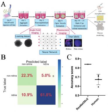
The three-dimensional, "organoid" approach for the differentiation of pluripotent stem cells into retinal and other neural tissues has become a major in vitro strategy to recapitulate development. In part, that success is owed to the availability of fluorescent reporter cell lines that allow to quickly and non-invasively assess differentiation. However, design and validation of reporter cell lines with genetic background of interest is time consuming and not compatible with clinical application. We hypothesized that basic contrast brightfield images contain sufficient information on the tissue specification and it is possible to extract this data using convolutional neural networks (CNN).Retina-specific reporter Rx-GFP mouse embryonic reporter stem cells have been used for the differentiation experiments. The brightfield (BF) images of organoids have been taken on day 6 and fluorescent on  day 9. To train the CNN we utilized a transfer learning approach: ImageNet pre-trained ResNet50v2 CNN had been trained on 3 000 labeled BF images, divided into two categories (retina and non-retina), based on the fluorescent reporter gene expression. A comparison of CNN with the human-based classifier showed that the CNN algorithm performs better than the expert in predicting organoid fate: 0.84% vs 0.67 ± 0.06% of correct predictions respectively, confirming our original hypothesis. Overall, we have demonstrated that computer algorithm can successfully predict retinal differentiation in organoids before the onset of reporter gene expression, which forms the basis for universal, non-invasive, scalable and rapid approach to assess the state of the cell and forecast its fate.

HSCI Retreat 2020 (Abstract 8, Figure 1)

Figure 1

Click here for larger figure

Figure 1. Convolutional neural networks are able to predict retinal differentiation in three-dimensional retinal organoids
(A) Experimental outline: organoids were imaged on day 6 using bright-field and on day 9 using fluorescent microscopy. Fluorescent images were used to assign true labels and bright-field ones for feeding neural network. (B) Confusion matrix for selected CNN – ResNet50v2. Values in the squares represent percentages of true negative, false positive, false negative and true positive predictions. Color LUT shows absolute number of images in each group. (C) Comparison for human-based classifier and CNN-based. CNN showed better results on classification task showing 0.84 accuracy score vs 0.67 ± 0.06 for human.

(Adapted from Kegeles et al., Front. Cell. Neurosci., 2020, 14, DOI: 10.3389/fncel.2020.00171)